Membrane structure
Membrane structure
Phospholipid bilayers
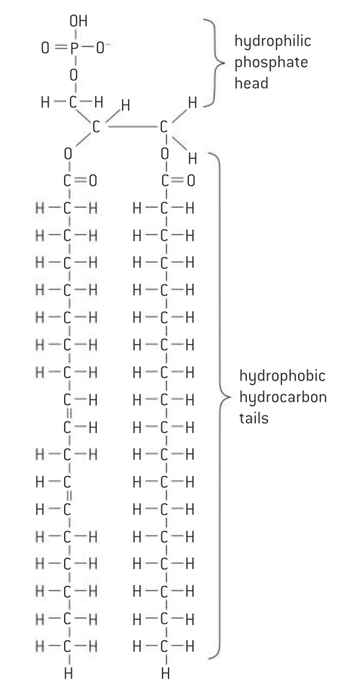
Phospholipids form bilayers in water due to the amphipathic properties of phospholipid molecules
- molecular structure of a phospholipid
-
important to emulate the degree of fluidity
Though it is difficult to determine whether the membrane is truly either a solid or liquid it can definitely be said to be fluid.
- Membranes need to be fluid enough that the cell can move
- Membranes need to be fluid enough that the required substances can move across the membrane
- If too fluid however the membrane could not effectively restrict the movement of substances across itself
-
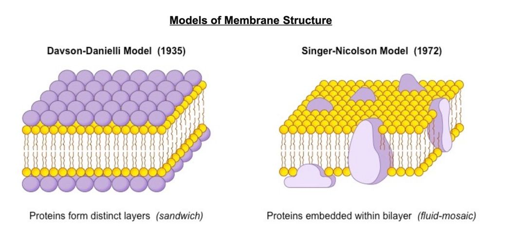
Davson and Danielli model 1930s
The evidence: In high magnification electron micrographs membranes appeared as two dark parallel lines with a lighter coloured region in between. Proteins appear dark in electron micrographs and phospholipids appear light - possibly indicating proteins layers either side of a phospholipid core.
The model:
- A protein-lipid sandwich
- Lipid bilayer composed of phospholipids(hydrophobic tails inside, hydrophilic heads outside)
- Proteins coat outer surface
- Proteins do not permeate the lipid bilayer
Problems with model:
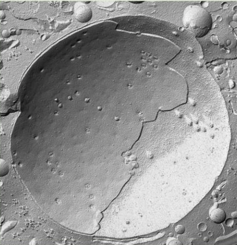
Freeze-etched electron micrographs
- this technique involves rapid freezing of cells and then fracturing them
- The fracture occurs along lines of weakness, including the centre of membranes.
- The fracture reveals an irregular rough surface inside the phospholipid bilayer
- structures of proteins being extracted later, they figure the varied in size can’t be what sandwich model said form plate layers
- Fluorescente antibody tagging
- The globular structures were interpreted as trans-membrane proteins.
Membrane Proteins
Membrane proteins are diverse in terms of structure, position in the membrane and function
- Functions of membrane proteins
- Hormone binding sites (receptors)
- Immobilized enzymes with the active site on the outside
- cell adhesion to form tight junctions between groups of cells in tissues and organs
- Cell-to-cell communication
- Channels for passive transport to allow hydrophilic particles across by facilitated diffusion
- Pumps for active transport which use ATP two move particles across the membrane
Integral protein
- hydrophobic on at least part of their surface
- embedded in the hydrocarbon chains in the centre of membrane
- usually extend across the membrane
Peripheral proteins
-
hydrophilic on their surface so are not embedded in membrane
-
mostly attached to surface of integral proteins
-
attachment is often reversible
-
types of protein
-
Receptor proteins: each binds to a specific molecule outside the cell which triggers a change inside the cell. Can be integral/peripheral (insulin receptor protein)
-
Enzyme proteins: promote chemical reactions that synthesize or break apart biological molecules. Can be integral/peripheral (ATP Synthase)
-
Adhesion proteins: Anchors the cell membrane to the inner cytoskeleton or proteins outside the cell as well as to other cells. Can be integral/peripheral (Cadherins, presence of calcium binds cells within tissues together)
-
Recognition proteins: Serve as identification tags on the surface of a cell. Often times these are glycoproteins (proteins with an attached sugar molecule) Can be integral/peripheral (MHC)
-
Glycoproteins
Protein produced by ribosome on RER—> add carbohydrates on protein to form glycoprotein/add fatty acid form lipoproteins by Golgi apparatus
- Carbohydrate groups may help position or orientate glycoproteins in membrane (prevent rotation in membrane)
- Carbohydrate groups may act as markers that determine the destination of a glycoprotein within the cell or for export (it may be removed after the protein has reached its destination)
- Important in intercellular recognition, interaction of different cells to form tissues, and detection of foreign cells by immune system
-
Channel proteins: Serve as pores/tunnels for large or hydrophilic molecules to be passively (no energy required) transported into/out of the membrane. All are integral
-
Pump Protein: Serve as pores/tunnels for large or hydrophilic molecules to be actively (energy required) transported into/out of the membrane. All are integral
-
Cholesterol in membranes
Cholesterol is a component of animal cell membranes
- structure of cholesterol
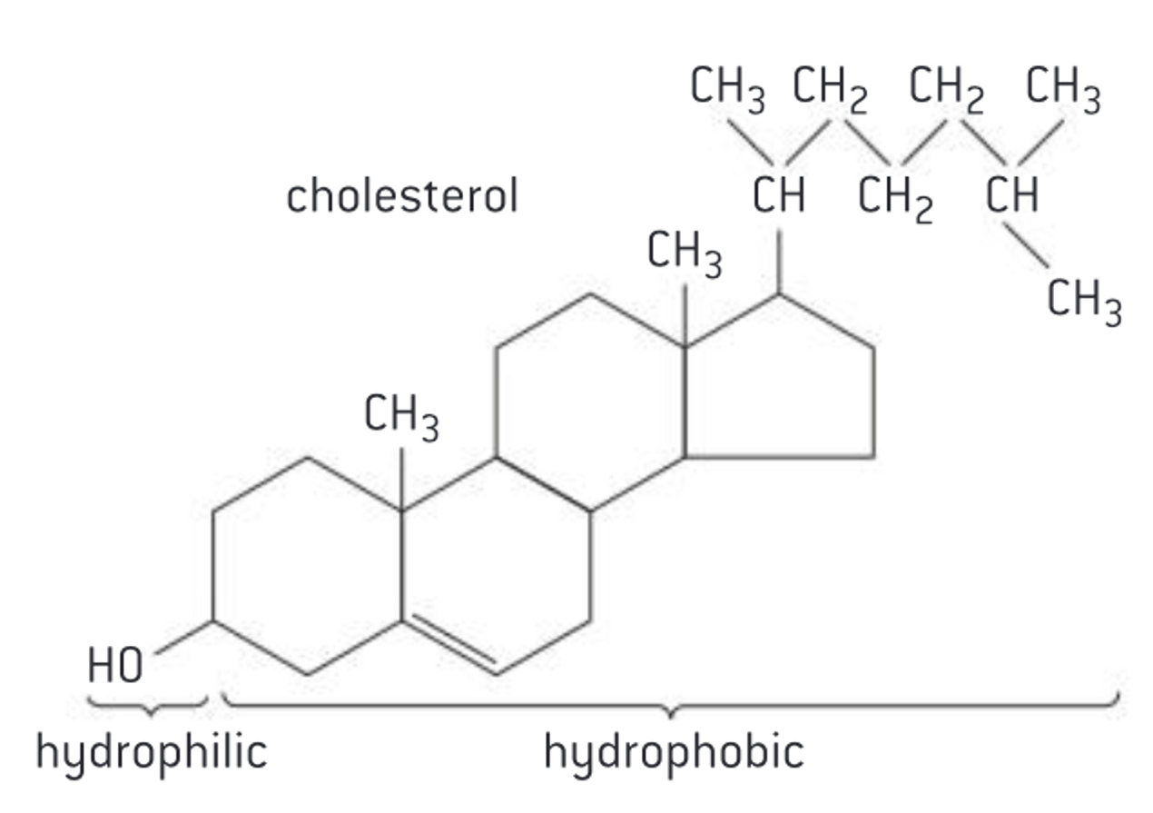
- type of steroids, liquid
- most of molecule is hydrophobic so attracted to hydrophobic hydrocarbon tails, but contain hydroxyl group (—OH) which is hydrophilic
- Hydrophilic group attracted to phosphate heads
Role of cholesterol in membranes
Cholesterol in mammalian membranes reduces membrane fluidity and permeability to some solutes
- hydrocarbon tails usually behave as liquid, phosphate heads act more like solid. Cell membranes do not correspond exactly to any of the 3 states.
- Overall membrane is fluid as components of the membrane are free to move
- Fluidity need to be control in membrane, too fluid—> less able to control what substances pass through, less fluid—> movement of cell and substances within it would be restricted
- Cholesterol disrupts regular packing of the hydro carbon tails, prevents them crystallizing and behaving as a solid
- It also restricts molecular motion and therefore fluidity of the membrane
- It also reduces the permeability to hydrophilic particles e.g. Na+
- Due to its shape cholesterol can help membranes to curve into a concave shape, which helps in the formation of vesicles during endocytosis